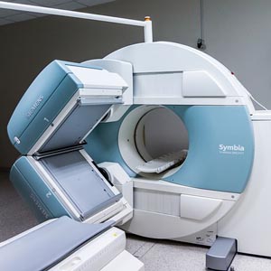
0.35 Tesla magnetic resonance imaging findings in a cohort of 399 seizure patients. Experience from a single centre in Nigeria

Accepted: 7 February 2022
All claims expressed in this article are solely those of the authors and do not necessarily represent those of their affiliated organizations, or those of the publisher, the editors and the reviewers. Any product that may be evaluated in this article or claim that may be made by its manufacturer is not guaranteed or endorsed by the publisher.
Authors
Epilepsy/seizures are major indications for brain imaging in clinical neurology. Structural lesions that may cause seizures are numerous and are defined using various neuroimaging techniques, including magnetic resonance imaging. The resolution of MRI allows for better fine ultra-structural lesions delineation. The aim of this study was to describe the pattern and frequency of structural brain lesions in MRI of patients with seizures and no clinically evident focal neurological signs. This was a retrospective, descriptive study carried out in a private hospital in Enugu, South East Nigeria to review all MRI results of patients who presented with seizures without clinical evidence of focal neurologic deficits. The MRI reports of two-third of the patients (47.9%) revealed focal lesions and about a third of the patients (32.2%) had normal findings. The structural lesions reported were mostly brain tumors (16%), stroke (9.5%), central nervous system infections (6.5%), brain malformation (6%) and encephalomalacia/gliosis (5%). Frequency of focal lesions clearly increased with age. Young patients were mostly associated with normal findings. Brain tumors and stroke were noted to occur more in the middle and aged patients respectively. Brain Magnetic Resonance Imaging remains a useful tool in the workup of patients with seizures without neurologic deficits. Treatable lesions can easily be revealed using this imaging modality.
How to Cite
PAGEPress has chosen to apply the Creative Commons Attribution NonCommercial 4.0 International License (CC BY-NC 4.0) to all manuscripts to be published.

 https://doi.org/10.4081/acbr.2022.188


