Computed tomographic pattern of stroke among adult patients in north-eastern Nigeria

Submitted: 23 April 2019
Accepted: 19 January 2020
Published: 14 July 2021
Abstract Views: 405
PDF: 167
Publisher's note
All claims expressed in this article are solely those of the authors and do not necessarily represent those of their affiliated organizations, or those of the publisher, the editors and the reviewers. Any product that may be evaluated in this article or claim that may be made by its manufacturer is not guaranteed or endorsed by the publisher.

Authors

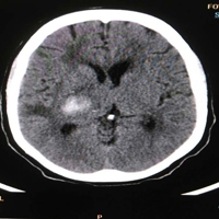
Stroke and its complications are major health problems in developing countries including Nigeria. It could be a major cause of death or disability especially when only clinical assessment is relied upon for diagnosis. Computed Tomography (CT) is a valuable tool for the diagnosis of stroke. CT pattern of stroke in the North Eastern Nigeria has not been fully described. This was a prospective descriptive study conducted at the Federal Teaching Hospital, Gombe, Nigeria from June 2016 to December 2016. One hundred and eleven patients who presented with clinical features of stroke and were referred to Radiology department for cranial CT were consecutively selected. Data were analysed using SPSS version 16.0 package. A p-value of ≤ 0.05 and confidence interval of 95% were adapted for statistical analysis. The variables were expressed as range, percentage and mean plus standard deviation. All comparison of variables was done applying kappa statistic and point-biserial correlation coefficient for the correlation analysis. There were 69 (62.2%) males and 42 (37.8%) females aged 18-90 years (mean ± SD of 57.49±13.47 years). Ninety-four (94) patients (84.7%) had ischaemic stroke, while the remaining 17 (15.3%) had haemorrhagic stroke. Lobar location was identified as the most common site of ischaemic stroke while thalamo-ganglionic area was the commonest location for haemorrhagic stroke. Age and hypertension were found to be the commonest risk factors associated with stroke. It is evident from this study that ischaemic stroke is the most prevalent stroke subtype. The middle cerebral artery territory was the commonest vascular territory involved in stroke while hypertension and age are common risk factors for both ischaemic and haemorrhagic stroke.

Dimensions

Altmetric

PlumX Metrics

Downloads

Download data is not yet available.

Citations

Wolf PA, Kannel WB, Dawber TR. Prospective investigations: The framingham study and the epidemiology of stroke. Adv Neurol 1977;19:107-20.
The World Health Organization MONICA Project (monitoring trends and determinants in cardiovascular disease): a major international collaboration. WHO MONICA Project Principal Investigators. J Clin Epidemiol 1988;41:105-14. DOI: https://doi.org/10.1016/0895-4356(88)90084-4
Donnan GA, Fisher M, Macleod M, Davis SM. Stroke. Lancet 2008;371:1612–23. DOI: https://doi.org/10.1016/S0140-6736(08)60694-7
Feigin VL. Stroke epidemiology in the developing world. Lancet 2005;365:2160–1. DOI: https://doi.org/10.1016/S0140-6736(05)66755-4
Nwosu MC. Epidemiology of stroke - an overview. J Med Invest Pract 2001;3:14-22.
Wahab KW. The burden of stroke in Nigeria. Int J Stroke 2008;3:290-2. DOI: https://doi.org/10.1111/j.1747-4949.2008.00217.x
Henon H, Godefroy O, Leys D, et al. Early predictors of death and disability after acute cerebral ischaemic event. Stroke 1995;26:292-8. DOI: https://doi.org/10.1161/01.STR.26.3.392
Ong TZ, Raymond AA. Risk factors for stroke and predictors of one-month mortality. Singapore Med J 2002;43:517-21.
Johnston KC, Li JY, Lyden PD, et al. Medical and neurological complications of ischaemic stroke. Experience from RANTTAS trial stroke 1998;29:447-53. DOI: https://doi.org/10.1161/01.STR.29.2.447
Eze U, Okaro O, Ohagwu C. Pattern of computed tomography findings in CVA patients in South-Eastern Nigeria. Eur J Sci Res 2009;34:104-9.
Mullin ME. Modern emergent stroke Imaging: Pearls, protocols and pitfalls. Radiol Clin N Am 2006;44:41-62. DOI: https://doi.org/10.1016/j.rcl.2005.08.002
Adams HP Jr, del Zoppo G, Alberts MJ, et al. Guidelines for the early management of adults with ischemic stroke: a guideline from the American Heart Association/American Stroke Association Stroke Council, Clinical Cardiology Council, Cardiovascular Radiology and Intervention Council, and the Atherosclerotic Peripheral Vascular Disease and Quality of Care Outcomes in Research Interdisciplinary Working Groups: the American Academy of Neurology affirms the value of this guideline as an educational tool for neurologists. Stroke 2007;38:1655-711. DOI: https://doi.org/10.1161/STROKEAHA.107.181486
de Lucas EM, Sánchez E, Gutiérrez A, et al. CT protocol for acute stroke: Tips and tricks for general radiologists. Radiographics 2008;28:1673-87. DOI: https://doi.org/10.1148/rg.286085502
Adeyekun AA, Ogunrin AO, Irabor PF. Computed tomographic predictors of mortality in haemorrhagic stroke. Trop J Health Sci 2007;14:13-8. DOI: https://doi.org/10.4314/tjhc.v14i2.36734
Chhetri K, Raut S. Computed tomography scan in the evaluation of patients with stroke. JCMS Nepal 2012;8:24-31. DOI: https://doi.org/10.3126/jcmsn.v8i2.6834
Kehinde O, Shamsideen AO, Mustapha AD, et al. Validation study of the siriraj stroke score in African Nigerians and evaluation of the discriminant values of its parameters. Stroke 2006;37:1997-2000. DOI: https://doi.org/10.1161/01.STR.0000229893.02732.02
Saad Al, Rajeh MD, Awada A, et al. Stroke in a Saudi Arabian National Guard community. Analysis of 500 consecutive cases from a population-based hospital. Stroke 1993;24:1635-9. DOI: https://doi.org/10.1161/01.STR.24.11.1635
Alkali HN, Sunday AB, Akano AO, et al. Stroke risk factors, subtypes, and 30‑day case fatality in Abuja, Nigeria. Niger Med J 2013;54:129-35. DOI: https://doi.org/10.4103/0300-1652.110051
Watila MM, Ibrahim A, Balarabe SA, et al. Risk factor profile among black stroke patients in Northeastern Nigeria. J Neurosci Behav Health 2012;31:50-8. DOI: https://doi.org/10.5897/JNBH11.052
Ukoha OB, Ajaegbu O, Eke CO. A review of stroke cases in a military hospital in Nigeria. Afrimedic J 2012;3:31-3.
Luntsi G, Pindiga BY, Ahmadu MS, et al. Evaluation of pattern of lesions depicted on brain Computed Tomography (CT) of patients presenting with stroke in a tertiary hospital in Northern Nigeria. IJAR 2015;1216-22.
Bwala, S. A., Stroke in a subsaharan Nigerian hospital—a retrospective study. Tropical Doctor, 1989;19(1): 11-14. DOI: https://doi.org/10.1177/004947558901900104
Chiewvit P, Danchaivijitr N, Nilanont Y, Poungvarin N. Computed tomographic findings in non-traumatic hemorrhagic stroke. J Med Assoc Thai 2009;92:73-5.
Robbins SL, Kumar V, Abbas AK. Robbins & Cotran Pathologic Basis of Disease. 9th Ed. Philadelphia, B. Saunders Company; 2010: pp. 1265-90.
Ikpeme AA, Bassey DE, Oku AO, Ephraim PE. Computerized tomography findings of cerebrovascular disease in adults in Calabar, Nigeria. West Afr J Radiol 2014;21:12-8. DOI: https://doi.org/10.4103/1115-1474.128074
Obiako OR, Oparah SK, Ogunniyi A. Prognosis and outcome of acute stroke in the University College Hospital Ibadan, Nigeria. Nigerian J Clinical Pract 2011;14;359-62. DOI: https://doi.org/10.4103/1119-3077.86784
Abubakar SA, Sabir AA. Profile of stroke patients seen in a tertiary health care centre in Nigeria. Ann Nigerian Med 2013;7:55 DOI: https://doi.org/10.4103/0331-3131.133097

How to Cite

Dahiru M, Yunusa, Umar Umar H, Dahiru Aminu MC, Aminu U Usman, Suleiman T Sa’ad, Philip Oluleke Ibinaiye, and Yusuf Hadiza. 2021. “Computed Tomographic Pattern of Stroke Among Adult Patients in North-Eastern Nigeria”. Pyramid Journal of Medicine 4 (1). https://doi.org/10.4081/pjm.2021.50.